jan 12, 2000 - α7nAChR
Description:
Kynurenic acid inhibits the normal function of α7nAChR by acting as a potent non-competitive α7nAChR-antagonist (Hilmas et al, 2001). α7nAChR dysfunction leads to abnormal NMDAR/GABAA function and perturbation of glutamatergic and GABAergic neurotransmission. In vivo and in vitro models have previously shown that loss of α7nAChR leads to diminished synaptic NMDARs as well as altered synaptic NMDAR function in cortical pyramidal cells. In particular, D-serine responsive synaptic NMDARs were shown to be specifically diminished physiologically due to loss of α7nAChR , along with serine racemase (Lin et al, 2014).A study using in vitro cultured cortical neurons has also demonstrated that D-serine and SR colocalize with PSD-95 in the postsynaptic terminals of glutamatergic synapses and exogenous application of D-serine, but not glycine, enhanced SR interactions with PSD-95 and NMDARS in a postsynaptic protein complex and increased the amount of glutamatergic synapses. However, antagonist-blockage at the glycine site of NMDARs inhibited the increase in synapse number, demonstrating that D-serine effects are mediated through synaptic NMDARs (Lin et al, 2016).
PSD-95 promotes spine synapse formation through direct interaction with postsynaptic neuronal nitric oxide synthase (nNOS) and drives presynaptic and postsynaptic maturation of glutamatergic synapses (El-Husseini et al., 2000; Nikonenko et al., 2008).
Increased concentrations of KYNA in the PFC have also been shown to reduce GABAergic transmission through a presynaptic 7nAChR-dependent mechanism by which the inhibitory effect of KYNA blocked 7nAChR signaling (Flores-Barrera, 2017). The PFC contains intrinsic glutamatergic pyramidal neurons and GABAergic interneurons, which are connected by multiple signaling mechanisms (Tseng and O'Donnell, 2004; Povysheva et al., 2008; DeFelipe et al., 2013).
Inhibition
of NMDARs during early life causes a persistent decrease in number of parvalbuminimmunoreactive
cells without cell death (Powell et al., 2012), suggesting that disruption of
NMDAR-mediated signaling impairs maturation of these cells.
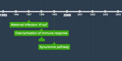
Interleukin-6 mediates ketamine-induced NOX upregulation and subsequently PVI deficits (Behrens et al., 2008). Early-life pro-inflammatory conditions, such as maternal and neonatal immune challenges, cause a persistent decrease in number of prefrontal and/or hippocampal parvalbumin-immunoreactive interneurons (Jenkins et al., 2009; Meyer et al., 2008). This deleterious effect could result from a redox dysregulation as maternal immune challenge transiently decreases GSH and vitamin E levels and increase oxidative stress in the hippocampus (Lante et al., 2007).
http://www.sciencedirect.com/science/article/pii/S0028390816302246#bib142
https://www.ncbi.nlm.nih.gov/pmc/articles/PMC5449450/#B151
https://academic.oup.com/endo/article-lookup/doi/10.1210/en.2011-0278
http://www.sciencedirect.com/science/article/pii/S0028390816303380
http://www.sciencedirect.com/science/article/pii/S0896627314000749
https://www.ncbi.nlm.nih.gov/pmc/articles/PMC3700678/
https://ac.els-cdn.com/S0166223612002044/1-s2.0-S0166223612002044-main.pdf?_tid=a8258260-a895-11e7-9dfb-00000aacb35f&acdnat=1507074818_3d2e9b3e8597409790e4b973424ecc7b
https://www.ncbi.nlm.nih.gov/pmc/articles/PMC3528177/
https://www.ncbi.nlm.nih.gov/pmc/articles/PMC2857580/
Added to timeline:
Date:
jan 12, 2000
Now
~ 25 years ago