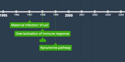
2 ene 1998 año - Over/activation of immune response
Descripción:
As a result of an infection, an immune response in triggered. Interferon-gamma (IFN-g) is a pleiotropic cytokine that is released by activated T-lymphocytes and natural killer cells and activates manys cells that originate from the immune system, including monocytes and macrophages (Senand Lengyel, 1992). Microglial are the resident macrophages of the CNS (Wake et al, 2009) and their activation triggers micro
glial polarization, which results in a microglia type 1 (M1) inflammatory state (Frank et al, 2007; Johnson et al, 2002). M1 microglia produce large amounts of inflammatory cytokines such as TNF, IL-1, IL-6, IL-12, and IL-1β (de Pablos et al, 2006; Blandino et al, 2006; Shimoda et al, 2006; Corin et al, 1996). Research has shown substantial increases in pro-inflammatory cytokine protein levels of TNFα, IL-1β, and IL-6 in maternal serum, amniotic fluid, and fetal brain following MIA (Oskvig, 2012). An unbalanced production of inflammatory cytokines affects neurite outgrowth, neuronal connections, and neurotransmitter formation, and induces neuronal cytotoxicity, all of which contribute to neuropsychiatric disorders (de Pablos et al, 2006; Mahadevan et al, 2017; Lee et al, 2017; Szymona et al, 2017). Correspondingly, postmortem and in vivo studies in schizophrenia patients have shown increased microglial density and microglial activation in the hippocampus and gray matter (Busse et al, 2012; Bayer et al, 1999; Radewicz et al, 2000; Steiner et al, 2006; Doorduin et al, 2009; van Berckel et al, 2008).
Microglial processes dynamically and preferentially interact with the dendritic spines of pyramidal neurons (Paolicelli et al., 2011; Tremblay et al., 2010; Wake et al., 2009). Furthermore, the processes of microglia that show the anatomical features that define an activated state have more frequent and prolonged contacts with, and increased phagocytosis of, dendritic spines (Tremblay et al., 2010). Complement components C3 and C4 are thought to mark synapses for phagocytosis by microglia expressing complement receptor 3 (CR3) (Paolicelli et al., 2011; Schafer et al., 2012; Sekar et al., 2016; Stevens et al., 2007). Taken together, these lines of evidence suggest that activated microglia could contribute to deficits in pyramidal neuron dendritic spines in the disorder.
Añadido al timeline:
New timeline
fecha:


